U.S. flag

An official website of the United States government

NCBI Bookshelf. A service of the National Library of Medicine, National Institutes of Health.

Adam MP, Feldman J, Mirzaa GM, et al., editors. GeneReviews® [Internet]. Seattle (WA): University of Washington, Seattle; 1993-2024.

Cover of GeneReviews®

GeneReviews® [Internet].

Show details

Ellis-van Creveld Syndrome

Synonym: Chondroectodermal Dysplasia (Ellis-van Creveld)

, MD, PhD, , MD, and , MD.

Author Information and Affiliations

Initial Posting: .

Estimated reading time: 26 minutes

Summary

Clinical characteristics.

Ellis-van Creveld (EVC) syndrome is characterized by postaxial polydactyly of the hands, disproportionate short stature with short limbs, dystrophic and/or hypoplastic nails, dental and oral manifestations, congenital heart disease, and radiologic abnormalities (narrow chest, short ribs, short tubular bones, bulbous ends of the proximal ulnae and distal radii, carpal and metacarpal fusions, cone-shaped epiphyses of phalanges, small iliac crests, acetabular spur projections [trident ilia], and lateral slanting of the tibial plateau). Other less common and more variable features include postaxial polydactyly of the feet, upper lip defect, and developmental delay.

Diagnosis/testing.

The diagnosis of EVC syndrome is established in a proband with characteristic clinical and radiographic findings and biallelic pathogenic variants in DYNC2H1, DYNC2LI1, EVC, EVC2, GLI, SMO, or WDR35 or a heterozygous pathogenic variant in PRKACA or PRKACB identified by molecular genetic testing.

Management.

Treatment of manifestations: Surgical amputation for polydactyly if desired; surgical correction of genu valgum as needed; physical therapy as needed; mechanical ventilation may be required in the neonatal period in those with severe restrictive lung disease; orthodontic and/or surgical treatment of dental anomalies; standard treatment for congenital heart disease; developmental services as needed; surgical correction of genital urinary malformations as needed; standard treatment for hearing loss.

Surveillance: Monitor growth at least annually throughout childhood; orthopedic evaluation with radiographic assessment as needed; physical and rehabilitation medicine evaluation as needed; assessment for manifestations of respiratory failure and/or restrictive lung disease as needed; monitor for dental eruption, overcrowding, and dental morphology annually throughout childhood; echocardiogram as needed; monitor developmental progress and educational needs at each visit until adulthood; hearing evaluation as needed if hearing loss is present.

Pregnancy management: Cesarean delivery is recommended in pregnant women with EVC syndrome who have pelvic/hip abnormalities.

Genetic counseling.

EVC syndrome caused by pathogenic variants in DYNC2H1, DYNC2LI1, EVC, EVC2, GLI, SMO, or WDR35 is inherited in an autosomal recessive manner. EVC syndrome caused by pathogenic variants in PRKACA or PRKACB (accounting for 2% of affected individuals) is inherited in an autosomal dominant manner.

Autosomal recessive inheritance: If both parents are known to be heterozygous for an EVC syndrome-related pathogenic variant, each sib of an affected individual has at conception a 25% chance of being affected, a 50% chance of being heterozygous, and a 25% chance of inheriting neither of the familial pathogenic variants. Once the EVC syndrome-related pathogenic variants have been identified in an affected family member, carrier testing for at-risk relatives and prenatal and preimplantation genetic testing are possible.

Diagnosis

No consensus clinical diagnostic criteria for Ellis-van Creveld (EVC) syndrome have been published.

Suggestive Findings

EVC syndrome should be suspected in probands with a combination of the following clinical and imaging findings and family history.

Clinical findings

  • Bilateral postaxial polydactyly of the hands (see Figure 1B-C) with or without postaxial polydactyly of the feet
  • Limb shortening (prenatal or postnatal)
  • Disproportionate short stature (prenatal or postnatal onset)
  • Dystrophic and/or hypoplastic nails (See Figure 1B-E.)
  • Dental and oral anomalies (hypodontia, delayed eruption of teeth, frenulum abnormalities)
  • Congenital heart defects (atrial septal defect, ventricular septal defect, single atrium, atrioventricular canal)
Figure 1.

Figure 1.

Clinical and radiographic images of a male child with Ellis-van Creveld (EVC) syndrome A. Dysmorphic features at age 19 months with short broad nose and partial upper lip defect in the midline.

Imaging findings (See Figure 1F-K.)

  • Narrow chest with short ribs
  • Short and thickened tubular bones
  • Bulbous ends of the proximal ulnae and distal radii
  • Carpal and metacarpal fusions (typically capitate and hamate)
  • Cone-shaped epiphyses of phalanges
  • Small iliac crests
  • Acetabular spur projections (trident ilia)
  • Lateral slanting of the tibial plateau

Family history is consistent with autosomal recessive inheritance (e.g., affected sibs and/or parental consanguinity). Absence of a known family history does not preclude the diagnosis.

Establishing the Diagnosis

The diagnosis of EVC syndrome is established in a proband with suggestive findings and biallelic pathogenic (or likely pathogenic) variants in DYNC2H1, DYNC2LI1, EVC, EVC2, GLI, SMO, or WDR35 or a heterozygous pathogenic (or likely pathogenic) variant in PRKACA or PRKACB identified by molecular genetic testing.

Note: (1) Per ACMG/AMP variant interpretation guidelines, the terms "pathogenic variant" and "likely pathogenic variant" are synonymous in a clinical setting, meaning that both are considered diagnostic and can be used for clinical decision making [Richards et al 2015]. Reference to "pathogenic variants" in this section is understood to include any likely pathogenic variants. (2) The identification of variant(s) of uncertain significance cannot be used to confirm or rule out the diagnosis.

Molecular genetic testing approaches can include a combination of gene-targeted testing (multigene panel) and comprehensive genomic testing (exome sequencing, genome sequencing). Gene-targeted testing requires that the clinician determine which gene(s) are likely involved (see Option 1), whereas comprehensive genomic testing does not (see Option 2).

Option 1

A skeletal dysplasia multigene panel that includes some or all of the genes listed in Table 1 and other genes of interest (see Differential Diagnosis) is most likely to identify the genetic cause of the condition while limiting identification of variants of uncertain significance and pathogenic variants in genes that do not explain the underlying phenotype. Note: (1) The genes included in the panel and the diagnostic sensitivity of the testing used for each gene vary by laboratory and are likely to change over time. (2) Some multigene panels may include genes not associated with the condition discussed in this GeneReview. (3) In some laboratories, panel options may include a custom laboratory-designed panel and/or custom phenotype-focused exome analysis that includes genes specified by the clinician. (4) Methods used in a panel may include sequence analysis, deletion/duplication analysis, and/or other non-sequencing-based tests.

For an introduction to multigene panels click here. More detailed information for clinicians ordering genetic tests can be found here.

Note: Targeted analysis for EVC founder variant c.1886+5G>T can be performed first in individuals of Amish ancestry (see Table 9).

Option 2

When the phenotype is indistinguishable from many other inherited disorders characterized by skeletal and/or ectodermal dysplasia, comprehensive genomic testing does not require the clinician to determine which gene(s) are likely involved. Exome sequencing is most commonly used; genome sequencing is also possible.

For an introduction to comprehensive genomic testing click here. More detailed information for clinicians ordering genomic testing can be found here.

Table 1.

Molecular Genetic Testing Used in Ellis-van Creveld Syndrome

Gene 1, 2Proportion of EVC Syndrome Attributed to Pathogenic Variants in GeneProportion of Pathogenic Variants 3 Detectable by Method
Sequence analysis 4Gene-targeted deletion/duplication analysis 5
DYNC2H1 2% 6100% 6None reported 7
DYNC2LI1 3% 8100% 8None reported 7
EVC (EVC1)49% 984% 916% 9, 10
EVC2 38% 990% 910% 9, 10
GLI1 4% 11100% 11None reported 7
PRKACA 1% 12100% 12None reported 7
PRKACB 1% 13100% 13None reported 7
SMO <1% 14100% 14None reported 7
WDR35 2% 15100% 15None reported 7

EVC = Ellis-van Creveld

1.

Genes are listed in alphabetic order.

2.
3.

See Molecular Genetics for information on variants detected in these genes.

4.

Sequence analysis detects variants that are benign, likely benign, of uncertain significance, likely pathogenic, or pathogenic. Variants may include missense, nonsense, and splice site variants and small intragenic deletions/insertions; typically, exon or whole-gene deletions/duplications are not detected. For issues to consider in interpretation of sequence analysis results, click here.

5.

Gene-targeted deletion/duplication analysis detects intragenic deletions or duplications. Methods used may include a range of techniques such as quantitative PCR, long-range PCR, multiplex ligation-dependent probe amplification (MLPA), and a gene-targeted microarray designed to detect single-exon deletions or duplications. Exome and genome sequencing may be able to detect deletions/duplications using breakpoint detection or read depth; however, sensitivity can be lower than gene-targeted deletion/duplication analysis.

6.
7.

Data derived from the subscription-based professional view of Human Gene Mutation Database [Stenson et al 2020]

8.
9.
10.

Approximately 23% of reported pathogenic copy number variants predictably affect both EVC and EVC2, as their coding sequences lie contiguously in the genome.

11.
12.
13.

Palencia-Campos et al [2020], Aubert-Mucca et al [2023]. One mosaic variant has been reported in one individual.

14.
15.

Clinical Characteristics

Clinical Description

Ellis-van Creveld (EVC) syndrome is characterized by postaxial polydactyly of the hands, disproportionate short stature, features of ectodermal dysplasia, congenital heart disease, and radiologic abnormalities (such as short ribs and short tubular bones) [Da Silva et al 2023]. Other less common and more variable features include postaxial polydactyly of the feet, nonspecific dysmorphic facial features, and developmental delay. To date, approximately 250 individuals with EVC syndrome have been identified with pathogenic variants in one of the genes listed in Table 1 [Aubert-Mucca et al 2023, Da Silva et al 2023]. The following description of the phenotypic features associated with this condition is based on these reports.

Table 2.

Ellis-van Creveld Syndrome: Frequency of Select Features

Feature% of Persons w/FeatureComment
Postaxial polydactyly (hands) 98%Bilateral in >95%
Limb shortening 83%Prenatal limb shortening in 36%
Nail dystrophy/hypoplasia 78%
Short stature 73%
Congenital heart disease 66%Atrial septal defect in >80%
Thoracic narrowing 66%Typically symmetric
Dental anomalies 59%
Brachydactyly 35%
Postaxial polydactyly (feet) 34%Bilateral in >90%
Upper lip defect 28%
Developmental delay 9%

Growth deficiency. Disproportionate short stature is common in individuals with EVC syndrome. About one third of individuals manifest growth deficiency in the perinatal setting, with shortened long bones evident on prenatal ultrasound, as well as short stature and/or limb shortening at birth [Da Silva et al 2023]. Most children present with growth failure and consequent short stature. Body segments are disproportionate, with a long, narrow thorax, usually accompanied by shortening of all four limbs. Limb segments are typically affected, with increasing severity from proximal to distal (rhizomelia > mesomelia > acromelia) [Baujat & Le Merrer 2007]. Adult height is most often below the midparental height and ranges between 109 and 152 cm.

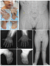
Musculoskeletal manifestations. The most common feature in individuals with EVC syndrome is bilateral postaxial polydactyly of the hands (see Figure 1B-C and Figure 1F-G) [Da Silva et al 2023]. Almost all individuals have hexadactyly of each hand, although a smaller subset have heptadactyly (seven digits) on each hand. Postaxial polydactyly of the feet is less common and is almost always bilateral. Polydactyly can sometimes be identified on prenatal ultrasound.

The thorax is usually long and narrow due to underdevelopment of the rib cage / short ribs, which may lead to lung hypoplasia during development or restrictive lung disease after birth. However, respiratory insufficiency is rarely severe [Huber & Cormier-Daire 2012]. Less commonly, pectus carinatum can be observed.

Other common skeletal abnormalities include shortening of long bones, brachydactyly (see Figure 1B-C and Figure 1F-I), carpal and metacarpal fusions (see Figure 1G), and genu valgum (due to a delay in ossification of the lateral tibial metaphysis) (see Figure 1K) [Handa et al 2020, Da Silva et al 2023]. The ulnae and radii usually have a distinct appearance due to the presence of abnormally bulbous ends. Bone age is usually delayed [Baujat & Le Merrer 2007]. Clinodactyly and syndactyly have also been reported.

The skull and spine are usually normal [Handa et al 2020]. Muscle strength is also normal.

Dental and oral manifestations. Anomalies of the teeth are frequent and varied. The most common findings are delayed eruption and hypodontia. Some individuals have natal teeth or early eruption of the primary dentition during the first two months of life and dental dysmorphism (e.g., cone-shaped teeth). Less frequent findings include enamel hypoplasia, dental transposition, and taurodontism [Peña-Cardelles et al 2019]. Frenulum anomalies, with multiple frenula and adhesions, are also common [Da Silva et al 2023].

Nail dystrophy. Nails are usually hypoplastic, and all nails show at least mild dystrophy, including discoloration, brittleness, pitting, ingrown nails, and deep-set nails (see Figure 1B-E).

Congenital heart defects are relatively frequent and can be detected prenatally. When present, they almost always include an atrial septal defect that can be isolated or coexist with other malformations. Ventricular septal defects, single atrium, and left superior vena cava are also rather common. Just under half of individuals with congenital heart defects develop some degree of heart failure [Hills et al 2011]. Other types of malformations (e.g., hypoplastic left ventricle, pulmonary valve stenosis/atresia, aortic coarctation) are rare [Hills et al 2011].

Craniofacial features are variable and nonspecific, and there is no specific facial gestalt for EVC syndrome. Upper lip defects (see Figure 1A) frequently manifest with partial cleft lip (without cleft palate); disruption of the superior gingivolabial groove due to adhesions is frequently seen with the lip defect. Additional reported facial features described in less than one in four individuals include hypertelorism, short broad nose (see Figure 1A), long philtrum, and postnatal microcephaly [Da Silva et al 2023].

Development. Intellectual disability is not typical of EVC syndrome. Individuals usually exhibit normal intelligence. A small percentage of individuals exhibit developmental delay and/or intellectual disability that is mostly mild or moderate [Öztürk et al 2021, Zaka et al 2021, Qian et al 2022]. Motor skills are most often affected, which may be secondary to the musculoskeletal abnormalities.

Other. Other rare features (described in <5% of individuals with EVC syndrome) include:

  • Genitourinary abnormalities (epispadias, hypospadias, cryptorchidism, hydrometrocolpos, kidney malformations, kidney cysts)
  • Central nervous system malformations (Dandy-Walker malformation, corpus callosum hypoplasia, cerebellar hypoplasia)
  • Sensorineural deafness

Phenotype Correlations by Gene

EVC2. A detailed assessment showed increased frequency of some manifestations in individuals with EVC2-related EVC syndrome compared to those with pathogenic variants in EVC [Da Silva et al 2023], but the phenotype in those with EVC-related EVC syndrome is most often clinically indistinguishable from that of EVC2-related EVC syndrome. Individuals with biallelic EVC2 pathogenic variants have increased shortening and thickening of the tubular bones and lower weight than individuals with EVC pathogenic variants. This suggests (but does not confirm) increased severity for EVC2-related EVC syndrome.

DYNC2H1, DYNC2LI1, GLI1, PRKACA, PRKACB, SMO, and WDR35. EVC syndrome due to pathogenic variants in DYNC2H1, DYNC2LI1, GLI1, PRKACA, PRKACB, SMO, or WDR35 are rare, so phenotype correlations by gene cannot be definitively established. However, postaxial polydactyly of the feet seems to be more common (and present in most individuals) with DYNC2H1-, DYNC2LI1-, GLI1-, SMO-, and WDR35-related EVC syndrome [Caparrós-Martín et al 2015, Palencia-Campos et al 2017, Niceta et al 2018, Aubert-Mucca et al 2023, Piceci-Sparascio et al 2023].

Genotype-Phenotype Correlations

EVC syndrome caused by large intragenic deletions or duplications affecting EVC and/or EVC2 are associated with more atypical clinical presentations, with a decrease in the proportion of musculoskeletal findings and an increase in the frequency of dysmorphic features [Da Silva et al 2023].

EVC

  • Missense variants are associated with decreased frequency of common skeletal findings (e.g., shortening and thickening of tubular bones, small iliac crest).
  • The coding region of EVC partially overlaps with the coding region of CRMP1. Individuals with pathogenic variants affecting the coding region shared between EVC and CRMP1 have an increased incidence of musculoskeletal and ectodermal dysplasia features compared to those with pathogenic variants only within the coding region of EVC. Therefore, CRMP1 has been suggested as a modifier of severity in individuals with EVC-related EVC syndrome [Da Silva et al 2023].

No definitive genotype-phenotype correlations have been identified for DYNC2H1-, DYNC2LI1-, GLI1-, PRKACA-, PRKACB-, SMO-, or WDR35-related EVC syndrome.

Nomenclature

In the 2023 revised Nosology of Genetic Skeletal Disorders [Unger et al 2023], EVC syndrome is included in Group 10, "Skeletal disorders caused by abnormalities of cilia or ciliary signaling," and referred to as "chondroectodermal dysplasia (Ellis-van Creveld)" followed by the involved gene – e.g., chondroectodermal dysplasia (Ellis-van Creveld), EVC2-related.

Prevalence

The exact prevalence of EVC syndrome is unknown. To date, there have been approximately 250 individuals reported with EVC syndrome and confirmed pathogenic variants identified in one of the genes listed in Table 1.

EVC syndrome may have an increased prevalence in individuals of Amish descent, as there is a founder EVC variant (c.1886+5G>T) in the Amish population of Lancaster County, Pennsylvania [McKusick 2000].

Differential Diagnosis

Table 4.

Skeletal Ciliopathies of Interest in the Differential Diagnosis of Ellis-van Creveld Syndrome

GenesDisorderSkeletal ManifestationsExtraskeletal ManifestationsComments
CEP120
CFAP410
DYNC2H1
DYNC2I1 (WDR60)
DYNC2I2 (WDR34)
DYNC2LI1
DYNLT2B (TCTEX1D2)
GRK2
IFT122
IFT140
IFT172
IFT43
IFT52
IFT80
IFT81
KIAA0586
KIAA0753
TRAF3IP1
TTC21B
WDR19
WDR35
Short-rib thoracic dysplasia (SRTD) (formerly asphyxiating thoracic dysplasia, or Jeune syndrome) 1, 2
  • Thoracic hypoplasia (wide spectrum of severity ranging from mild form to lethal condition)
  • Handlebar clavicles
  • Short trident pelvis/ilia
  • Short tubular bones
  • Brachydactyly
  • Cone-shaped epiphyses
  • Postaxial polydactyly (uncommon)
  • Retinal degeneration
  • Pulmonary hypoplasia
  • Cystic disease (liver, pancreas, kidney)
  • Kidney failure
  • Liver failure
  • Respiratory impairment (due to thoracic anomalies) is much more common & severe in SRTD than in EVC syndrome, being the hallmark of SRTD.
  • Kidney & liver are also commonly affected in SRTD.
  • Polydactyly is uncommon in SRTD.
DYNC2I1 (WDR60)
DYNC2I2 (WDR34)
DYNC2LI1
IFT122
IFT80
IFT81
INTU
NEK1
TRAF3IP1
WDR19
Short-rib polydactyly syndrome (SRPS) 1, 3
  • Severe thoracic hypoplasia
  • Polydactyly (axis variable)
  • Bulbous end of long bones
  • Bowed radii & ulnae
  • Tibial hypoplasia
  • Hydropic appearance
  • Lingual hamartomas
  • Bifid tongue
  • Malformations (heart, lung, epiglottis, kidney, pancreas, genitalia)
  • Imperforate anus
  • Holoprosencephaly
  • SRPS is typically much more severe than EVC syndrome, w/high rate of perinatal mortality.
  • Multiple malformations are common in SRPS.
  • Polydactyly is variable in SRPS.
EVC
EVC2
Weyers acrofacial (acrodental) dysostosis (WAD) 1, 4
  • Postaxial polydactyly
  • Mild short stature
  • Short hands w/mild brachydactyly
  • Nail dystrophy
  • Dental anomalies (conical teeth, hypodontia, delayed eruption)
  • Multiple frenula
  • WAD is considered a mild form of EVC syndrome (less prominent skeletal features).
  • Postaxial polydactyly of feet is more common in WAD than in EVC syndrome.
IFT122
IFT40
IFT43
WDR19
WDR35
Cranioectodermal dysplasia (CED) (Levin-Sensenbrenner) 1, 5
  • Craniosynostosis
  • Narrow thorax
  • Short proximal bones
  • Severe brachydactyly
  • Postaxial polydactyly (uncommon)
  • Ectodermal dysplasia
  • Characteristic facial features w/frontal bossing & low-set ears
  • Loose skin & joint laxity
  • Progressive kidney failure
  • Hepatic disease
  • Retinal dystrophy
  • Unlike EVC syndrome, CED is assoc w/craniosynostosis, kidney failure, hepatic disease, & retinal dystrophy.
  • Polydactyly is uncommon in CED.
IFT140
IFT172
WDR19
Mainzer-Saldino syndrome (MZSDS) 1, 6
  • Craniosynostosis
  • Short stature
  • Cone-shaped epiphyses of phalanges
  • Femoral dysplasia (small & flattened epiphyses, short neck)
  • Microcephaly
  • Dental anomalies
  • Nystagmus
  • Retinal pigmentary dystrophy
  • Progressive kidney failure
  • Hepatic disease (uncommon)
  • MZSDS is assoc w/a less pronounced skeletal phenotype.
  • Retinal dystrophy is invariably present in MZSDS.
  • Kidney failure & malformation are also very common in MZSDS.

EVC = Ellis-van Creveld; MZSDS = Mainzer-Saldino syndrome; SRPS = short-rib polydactyly syndrome; CED = cranioectodermal dysplasia; SRTD = short-rib thoracic dysplasia; WAD = Weyers acrofacial (acrodental) dysostosis

1.
2.
3.
4.
5.
6.

Management

No clinical practice guidelines for Ellis-van Creveld (EVC) syndrome have been published.

Evaluations Following Initial Diagnosis

To establish the extent of disease and needs in an individual diagnosed with EVC syndrome, the evaluations summarized in Table 5 (if not performed as part of the evaluation that led to the diagnosis) are recommended.

Table 5.

Ellis-van Creveld Syndrome: Recommended Evaluations Following Initial Diagnosis

System/ConcernEvaluationComment
Growth Measurement of height, weight, & head circumference
Musculoskeletal
  • Physical exam w/measurement of body & limb segments
  • Complete radiographic skeletal survey
  • Physical & rehab medicine eval to assess need for PT
Respiratory Assessment of respiratory failure &/or restrictive lung diseaseIn those w/manifestations of significant thoracic narrowing &/or respiratory distress
Dental Dental examTo assess for hypodontia & teeth anomalies that require treatment & for need to remove natal teeth
Cardiovascular Echocardiogram
Development Developmental assessment
Genitourinary Clinical exam for genital anomalies & renal/pelvic ultrasound
Neurologic Neurologic evalConsider brain MRI in those w/abnormal neurologic exam.
Audiologic Audiologic eval
Genetic counseling By genetics professionals 1To inform affected persons & their families re nature, MOI, & implications of EVC syndrome to facilitate medical & personal decision making
Family support
& resources
By clinicians, wider care team, & family support organizationsAssessment of family & social structure to determine need for:

EVC = Ellis-van Creveld; MOI = mode of inheritance; PT = physical therapy

1.

Medical geneticist, certified genetic counselor, certified advanced genetic nurse

Treatment of Manifestations

Supportive treatment to improve quality of life, maximize function, and reduce complications is recommended. This ideally involves multidisciplinary care by specialists in relevant fields (see Table 6).

Table 6.

Ellis-van Creveld Syndrome: Treatment of Manifestations

Manifestation/ConcernTreatmentConsiderations/Other
Polydactyly Surgical amputation (if desired)
Musculoskeletal Surgical correction of genu valgumIf required per orthopedic assessment
PTPer physical & rehab medicine
Respiratory failure Mechanical ventilationMay be required in neonatal period &/or in persons w/severe restrictive lung disease
Dental anomalies Orthodontic &/or surgical treatment of dental anomalies
Congenital heart disease Standard treatment per cardiologist/cardiac surgeon
Developmental delay Developmental services (PT &/or OT) as needed in those w/developmental delay
Genitourinary malformations Surgical correction (if indicated)
Hearing loss Standard treatment for hearing loss

PT = physical therapy; OT = occupational therapy

Surveillance

To monitor existing manifestations, the individual's response to supportive care, and the emergence of new manifestations, the evaluations summarized in Table 7 are recommended.

Table 7.

Ellis-van Creveld Syndrome: Recommended Surveillance

System/ConcernEvaluationFrequency
Growth Monitor length, weight, & head circumference.At least annually throughout childhood
Musculoskeletal Orthopedic eval w/radiographic assessmentAs needed
Physical & rehab medicine evalAs needed in those requiring PT
Respiratory Assess for manifestations of respiratory failure / restrictive lung disease.As needed, if signs/risks for respiratory failure are present
Dental Monitor for dental eruption, overcrowding, & dental morphology.Annually throughout childhood
Cardiovascular EchocardiogramAs needed
Development Monitor developmental progress & educational needs.At each visit, until adulthood
Audiologic Hearing evalAs needed, if hearing loss is present

PT = physical therapy

Evaluation of Relatives at Risk

See Genetic Counseling for issues related to testing of at-risk relatives for genetic counseling purposes.

Pregnancy Management

In pregnant women with EVC syndrome who have pelvic/hip abnormalities, cesarean delivery is recommended.

See MotherToBaby for further information on medication use during pregnancy.

Therapies Under Investigation

Search ClinicalTrials.gov in the US and EU Clinical Trials Register in Europe for access to information on clinical studies for a wide range of diseases and conditions. Note: There may not be clinical trials for this disorder.

Genetic Counseling

Genetic counseling is the process of providing individuals and families with information on the nature, mode(s) of inheritance, and implications of genetic disorders to help them make informed medical and personal decisions. The following section deals with genetic risk assessment and the use of family history and genetic testing to clarify genetic status for family members; it is not meant to address all personal, cultural, or ethical issues that may arise or to substitute for consultation with a genetics professional. —ED.

Mode of Inheritance

Ellis-van Creveld (EVC) syndrome caused by pathogenic variants in DYNC2H1, DYNC2LI1, EVC, EVC2, GLI, SMO, or WDR35 is inherited in an autosomal recessive manner.

EVC syndrome caused by pathogenic variants in PRKACA or PRKACB (accounting for 2% of affected individuals) is inherited in an autosomal dominant manner [Palencia-Campos et al 2020, Aubert-Mucca et al 2023]. Autosomal dominant inheritance is not discussed further in this section.

Risk to Family Members (Autosomal Recessive Inheritance)

Parents of a proband

  • The parents of an affected child are presumed to be heterozygous for an EVC syndrome-related pathogenic variant.
  • Molecular genetic testing is recommended for the parents of a proband to confirm that both parents are heterozygous for an EVC syndrome-related pathogenic variant and to allow reliable recurrence risk assessment.
  • If a pathogenic variant is detected in only one parent and parental identity testing has confirmed biological maternity and paternity, it is possible that one of the pathogenic variants identified in the proband occurred as a de novo event in the proband or as a postzygotic de novo event in a mosaic parent [Jónsson et al 2017]. If the proband appears to have homozygous pathogenic variants (i.e., the same two pathogenic variants), additional possibilities to consider include:
  • Heterozygotes (carriers) are usually asymptomatic and are not at risk of developing EVC syndrome. However, a small number of heterozygous variants (typically EVC2 truncating variants in exon 22) have been associated with Weyers acrofacial dysostosis (see Table 3) [Da Silva et al 2023].

Sibs of a proband

  • If both parents are known to be heterozygous for a pathogenic variant in one of the genes listed in Table 1, each sib of an affected individual has at conception a 25% chance of being affected, a 50% chance of being heterozygous, and a 25% chance of inheriting neither of the familial pathogenic variants.
  • Heterozygotes (carriers) are usually asymptomatic and are not at risk of developing EVC syndrome. However, a small number of heterozygous variants (typically EVC2 truncating variants in exon 22) have been associated with Weyers acrofacial dysostosis (see Table 3) [Da Silva et al 2023].

Offspring of a proband. Unless an affected individual's reproductive partner also has EVC syndrome or is heterozygous for an EVC syndrome-related pathogenic variant, offspring will be obligate heterozygotes (carriers) for an EVC syndrome-related pathogenic variant.

Other family members. Each sib of the proband's parents is at a 50% risk of being a carrier of a pathogenic variant of one of the genes listed in Table 1.

Carrier Detection

Carrier testing for at-risk relatives requires prior identification of the EVC syndrome-related pathogenic variants in the family.

Related Genetic Counseling Issues

Family planning

  • The optimal time for determination of genetic risk and discussion of the availability of prenatal/preimplantation genetic testing is before pregnancy.
  • It is appropriate to offer genetic counseling (including discussion of potential risks to offspring and reproductive options) to young adults who are affected, are carriers, or are at risk of being carriers.
  • Carrier testing for the reproductive partners of known carriers should be considered, particularly if both partners are of the same ancestry. An EVC founder variant has been identified in individuals of Amish ancestry (see Table 9).

Prenatal Testing and Preimplantation Genetic Testing

Molecular genetic testing. Once the EVC syndrome-related pathogenic variants have been identified in a family member with autosomal recessive EVC syndrome, prenatal and preimplantation genetic testing are possible.

Fetal ultrasound examination. In a fetus not already known to be at risk for EVC syndrome based on family history, the identification on fetal ultrasound examination of the following features may prompt consideration of gene-targeted testing for known EVC syndrome-related genes: shortening of long bones, polydactyly, thoracic narrowing, and congenital heart disease (typically endocardial cushion defects) (see Diagnosis) [Chen et al 2012]. Other findings that may be observed on ultrasound examination of an affected fetus include severe upper lip defects and genitourinary and central nervous system malformations (though the latter two features are rarely reported in EVC syndrome).

Differences in perspective may exist among medical professionals and within families regarding the use of prenatal testing. While most centers would consider use of prenatal testing to be a personal decision, discussion of these issues may be helpful.

Resources

GeneReviews staff has selected the following disease-specific and/or umbrella support organizations and/or registries for the benefit of individuals with this disorder and their families. GeneReviews is not responsible for the information provided by other organizations. For information on selection criteria, click here.

Molecular Genetics

Information in the Molecular Genetics and OMIM tables may differ from that elsewhere in the GeneReview: tables may contain more recent information. —ED.

Table B.

OMIM Entries for Ellis-van Creveld Syndrome (View All in OMIM)

165220GLI FAMILY ZINC FINGER 1; GLI1
176892PROTEIN KINASE, cAMP-DEPENDENT, CATALYTIC, BETA; PRKACB
225500ELLIS-VAN CREVELD SYNDROME; EVC
601500SMOOTHENED, FRIZZLED CLASS RECEPTOR; SMO
601639PROTEIN KINASE, cAMP-DEPENDENT, CATALYTIC, ALPHA; PRKACA
603297DYNEIN, CYTOPLASMIC 2, HEAVY CHAIN 1; DYNC2H1
604831EVC CILIARY COMPLEX SUBUNIT 1; EVC
607261EVC CILIARY COMPLEX SUBUNIT 2; EVC2
613602WD REPEAT-CONTAINING PROTEIN 35; WDR35
617083DYNEIN, CYTOPLASMIC 2, LIGHT INTERMEDIATE CHAIN 1; DYNC2LI1

Molecular Pathogenesis

EVC and EVC2 encode membrane proteins that form a complex in the basal body of primary cilia [Ruiz-Perez et al 2007]. They have an essential function as mediators of hedgehog signaling in primary cilia, which is one of the fundamental drivers of the development of the orofacial region, the endochondral plate of the axial skeleton, and cardiac morphogenesis [Louie et al 2020].

SMO encodes a transcription factor that is activated upon hedgehog binding, leading to the production of GLI proteins (such as that encoded by GLI1) [Tukachinsky et al 2010]. SMO also interacts with the EVC-EVC2 complex, which allows translocation of the GLI proteins to the tip of the primary cilia, which are the distal effectors of hedgehog signaling, modulating proliferation and differentiation of developing tissues [Louie et al 2020].

WDR35 encodes a protein that coats Golgi vesicles, functioning as a signal for transport into the primary cilia, affecting both assembly and cargo transport. Its disruption has been shown to impair the recruitment of EVC and SMO to the primary cilia [Caparrós-Martín et al 2015]. Similarly, DYNC2H1 and DYNC2LI1 encode for subunits of dynein-2, which has a key role in the transport of cargo to the primary cilia [Niceta et al 2018].

PRKACA and PRKACB encode subunits of the enzyme protein kinase A (PKA). PKA is an intracellular mediator of G protein-coupled receptors (GPCR) that is activated by increases in intracellular cAMP. In primary cilia PKA constitutively represses GLI proteins and, therefore, hedgehog signaling [Palencia-Campos et al 2020]. SMO activation upon hedgehog binding leads to removal of the cilia GPCR and, consequently, to PKA inactivation.

Mechanism of disease causation. The mechanism of disease for EVC and EVC2 is loss of function [Zhang et al 2016, Louie et al 2020]. Despite functioning together as a protein complex, they are nonredundant, and the loss of one protein is sufficient to impair the function of the complex. Less data is available regarding the mechanism of disease causation for the other genes associated with Ellis-van Creveld (EVC) syndrome. However, considering the mechanism of EVC and EVC2, as well as the known function of the other genes, it is likely that loss of function is the mechanism in DYNC2H1-, DYNC2LI1-, GLI1-, SMO-, and WDR35-related EVC syndrome. PRKACA- and PRKACB-related EVC syndrome may be due to gain of function, as causal variants have been shown to lead to PKA activity elevation due to increased cAMP sensitivity and, therefore, hedgehog signaling inhibition [Palencia-Campos et al 2020]. This is also in line with the autosomal dominant mode of inheritance for both genes.

Table 8.

Ellis-van Creveld Syndrome: Gene-Specific Laboratory Considerations

GeneSpecial Consideration
EVC While the coding regions of EVC & EVC2 lie contiguously in the genome, are arranged in divergent orientations, & are separated by 2.6 kb of genomic sequence, these genes share no homology w/each other or w/any other regions. 1
EVC2

Table 9.

Pathogenic Variants Referenced in This GeneReview by Gene

GeneReference SequencesDNA Nucleotide ChangePredicted Protein ChangeComment [Reference]
EVC NM_153717​.2 c.1886+5G>T--Founder variant in Amish population of Lancaster County, Pennsylvania [McKusick 2000]

Variants listed in the table have been provided by the authors. GeneReviews staff have not independently verified the classification of variants.

GeneReviews follows the standard naming conventions of the Human Genome Variation Society (varnomen​.hgvs.org). See Quick Reference for an explanation of nomenclature.

Chapter Notes

Author Notes

Jorge Diogo Da Silva, Nataliya Tkachenko, and Ana Rita Soares are actively involved in clinical research regarding individuals with Ellis-van Creveld (EVC) syndrome. They would be happy to communicate with persons who have any questions regarding diagnosis of EVC syndrome or other considerations.

The authors are also interested in hearing from clinicians treating families affected by EVC syndrome in whom no causative variant has been identified through molecular genetic testing of the genes known to be involved in this group of disorders.

Contact Dr Jorge Diogo Da Silva (moc.liamg@avlis.rcd.egroj) to inquire about the above subjects, or about the review of EVC or EVC2 variants of uncertain significance.

Acknowledgments

The authors would like to acknowledge Dr Catarina Martins Silva for her valuable help regarding the photographic records of the individual with EVC syndrome presented in Figure 1.

Revision History

  • 26 October 2023 (sw) Review posted live
  • 5 July 2023 (jds) Original submission

References

Literature Cited

  • Aubert-Mucca M, Huber C, Baujat G, Michot C, Zarhrate M, Bras M, Boutaud L, Malan V, Attie-Bitach T, Cormier-Daire V, et al. Ellis-van Creveld syndrome: clinical and molecular analysis of 50 Individuals. J Med Genet. 2023;60:337-45. [PubMed: 35927022]
  • Baujat G, Le Merrer M. Ellis-van Creveld syndrome. Orphanet J Rare Dis. 2007;2:27. [PMC free article: PMC1891277] [PubMed: 17547743]
  • Caparrós-Martín JA, De Luca A, Cartault F, Aglan M, Temtamy S, Otaify GA, Mehrez M, Valencia M, Vázquez L, Alessandri JL, Nevado J, Rueda-Arenas I, Heath KE, Digilio MC, Dallapiccola B, Goodship JA, Mill P, Lapunzina P, Ruiz-Perez VL. Specific variants in WDR35 cause a distinctive form of Ellis-van Creveld syndrome by disrupting the recruitment of the EvC complex and SMO into the cilium. Hum Mol Genet. 2015;24:4126-37. [PMC free article: PMC4560068] [PubMed: 25908617]
  • Chen CP, Chen CY, Chern SR, Su JW, Wang W. First-trimester prenatal diagnosis of Ellis-van Creveld syndrome. Taiwan J Obstet Gynecol. 2012;51:643-8. [PubMed: 23276573]
  • Da Silva JD, Soares AR, Fortuna AM, Tkachenko N. Establishing an objective clinical spectrum, genotype-phenotype correlations and CRMP1 as a modifier in the Ellis-van Creveld syndrome: the first systematic review of EVC and EVC2-associated conditions. Genet Med Open. 2023;1:100781.
  • Handa A, Voss U, Hammarsjö A, Grigelioniene G, Nishimura G. Skeletal ciliopathies: a pattern recognition approach. Jpn J Radiol. 2020;38:193-206. [PubMed: 31965514]
  • Hills CB, Kochilas L, Schimmenti LA, Moller JH. Ellis-van Creveld syndrome and congenital heart defects: presentation of an additional 32 cases. Pediatr Cardiol. 2011;32:977-82. [PubMed: 21533779]
  • Huber C, Cormier-Daire V. Ciliary disorder of the skeleton. Am J Med Genet C Semin Med Genet. 2012;160C:165-74. [PubMed: 22791528]
  • Jónsson H, Sulem P, Kehr B, Kristmundsdottir S, Zink F, Hjartarson E, Hardarson MT, Hjorleifsson KE, Eggertsson HP, Gudjonsson SA, Ward LD, Arnadottir GA, Helgason EA, Helgason H, Gylfason A, Jonasdottir A, Jonasdottir A, Rafnar T, Frigge M, Stacey SN, Th Magnusson O, Thorsteinsdottir U, Masson G, Kong A, Halldorsson BV, Helgason A, Gudbjartsson DF, Stefansson K. Parental influence on human germline de novo mutations in 1,548 trios from Iceland. Nature. 2017;549:519-22. [PubMed: 28959963]
  • Lin AE, Traum AZ, Sahai I, Keppler-Noreuil K, Kukolich MK, Adam MP, Westra SJ, Arts HH. Sensenbrenner syndrome (cranioectodermal dysplasia): clinical and molecular analyses of 39 patients including two new patients. Am J Med Genet A. 2013;161A:2762-76. [PubMed: 24123776]
  • Louie KW, Mishina Y, Zhang H. Molecular and cellular pathogenesis of Ellis-van Creveld syndrome: lessons from targeted and natural mutations in animal models. J Dev Biol. 2020;8:25. [PMC free article: PMC7711556] [PubMed: 33050204]
  • McKusick VA. Ellis-van Creveld syndrome and the Amish. Nat Genet. 2000;24:203-4. [PubMed: 10700162]
  • Niceta M, Margiotti K, Digilio MC, Guida V, Bruselles A, Pizzi S, Ferraris A, Memo L, Laforgia N, Dentici ML, Consoli F, Torrente I, Ruiz-Perez VL, Dallapiccola B, Marino B, De Luca A, Tartaglia M. Biallelic mutations in DYNC2LI1 are a rare cause of Ellis-van Creveld syndrome. Clin Genet. 2018;93:632-9. [PubMed: 28857138]
  • Öztürk Ö, Bağış H, Bolu S, Çevik MÖ. Ellis-van Creveld syndrome novel pathogenic variant in the EVC2 gene a patient from Turkey. Clin Case Rep. 2021;9:1973-6. [PMC free article: PMC8077313] [PubMed: 33936625]
  • Palencia-Campos A, Aoto PC, Machal EMF, Rivera-Barahona A, Soto-Bielicka P, Bertinetti D, Baker B, Vu L, Piceci-Sparascio F, Torrente I, Boudin E, Peeters S, Van Hul W, Huber C, Bonneau D, Hildebrand MS, Coleman M, Bahlo M, Bennett MF, Schneider AL, Scheffer IE, Kibæk M, Kristiansen BS, Issa MY, Mehrez MI, Ismail S, Tenorio J, Li G, Skålhegg BS, Otaify GA, Temtamy S, Aglan M, Jønch AE, De Luca A, Mortier G, Cormier-Daire V, Ziegler A, Wallis M, Lapunzina P, Herberg FW, Taylor SS, Ruiz-Perez VL. Germline and mosaic variants in PRKACA and PRKACB cause a multiple congenital malformation syndrome. Am J Hum Genet. 2020;107:977-88. [PMC free article: PMC7675002] [PubMed: 33058759]
  • Palencia-Campos A, Ullah A, Nevado J, Yildirim R, Unal E, Ciorraga M, Barruz P, Chico L, Piceci-Sparascio F, Guida V, De Luca A, Kayserili H, Ullah I, Burmeister M, Lapunzina P, Ahmad W, Morales AV, Ruiz-Perez VL. GLI1 inactivation is associated with developmental phenotypes overlapping with Ellis-van Creveld syndrome. Hum Mol Genet. 2017;26:4556-71. [PubMed: 28973407]
  • Peña-Cardelles JF, Domínguez-Medina DA, Cano-Durán JA, Ortega-Concepción D, Cebrián JL. Oral manifestations of Ellis-van Creveld syndrome. A rare case report. J Clin Exp Dent. 2019;11:e290-e295. [PMC free article: PMC6461737] [PubMed: 31001402]
  • Perrault I, Saunier S, Hanein S, Filhol E, Bizet AA, Collins F, Salih MA, Gerber S, Delphin N, Bigot K, Orssaud C, Silva E, Baudouin V, Oud MM, Shannon N, Le Merrer M, Roche O, Pietrement C, Goumid J, Baumann C, Bole-Feysot C, Nitschke P, Zahrate M, Beales P, Arts HH, Munnich A, Kaplan J, Antignac C, Cormier-Daire V, Rozet JM. Mainzer-Saldino syndrome is a ciliopathy caused by IFT140 mutations. Am J Hum Genet. 2012;90:864-70. [PMC free article: PMC3376548] [PubMed: 22503633]
  • Piceci-Sparascio F, Micale L, Torres B, Guida V, Consoli F, Torrente I, Onori A, Frustaci E, D'Asdia MC, Petrizzelli F, Bernardini L, Mancini C, Soli F, Cocciadiferro D, Guadagnolo D, Mastromoro G, Putotto C, Fontana F, Brunetti-Pierri N, Novelli A, Pizzuti A, Marino B, Digilio MC, Mazza T, Dallapiccola B, Ruiz-Perez VL, Tartaglia M, Castori M, De Luca A. Clinical variability in DYNC2H1-related skeletal ciliopathies includes Ellis-van Creveld syndrome. Eur J Hum Genet. 2023;31:479-84. [PMC free article: PMC10133340] [PubMed: 36599940]
  • Qian Y, Wang X, Tang W, Zou C. Microdeletion of 4p16.2 in children: a case report and literature review. Case Rep Genet. 2022;2022:6253690. [PMC free article: PMC9013304] [PubMed: 35437470]
  • Richards S, Aziz N, Bale S, Bick D, Das S, Gastier-Foster J, Grody WW, Hegde M, Lyon E, Spector E, Voelkerding K, Rehm HL, et al. Standards and guidelines for the interpretation of sequence variants: a joint consensus recommendation of the American College of Medical Genetics and Genomics and the Association for Molecular Pathology. Genet Med. 2015;17:405-24. [PMC free article: PMC4544753] [PubMed: 25741868]
  • Ruiz-Perez VL, Blair HJ, Rodriguez-Andres ME, Blanco MJ, Wilson A, Liu YN, Miles C, Peters H, Goodship JA. Evc is a positive mediator of Ihh-regulated bone growth that localises at the base of chondrocyte cilia. Development. 2007;134:2903-12. [PubMed: 17660199]
  • Schmidts M, Frank V, Eisenberger T, Al Turki S, Bizet AA, Antony D, Rix S, Decker C, Bachmann N, Bald M, Vinke T, Toenshoff B, Di Donato N, Neuhann T, Hartley JL, Maher ER, Bogdanović R, Peco-Antić A, Mache C, Hurles ME, Joksić I, Guć-Šćekić M, Dobricic J, Brankovic-Magic M, Bolz HJ, Pazour GJ, Beales PL, Scambler PJ, Saunier S, Mitchison HM, Bergmann C. Combined NGS approaches identify mutations in the intraflagellar transport gene IFT140 in skeletal ciliopathies with early progressive kidney disease. Hum Mutat. 2013;34:714-24. [PMC free article: PMC4226634] [PubMed: 23418020]
  • Stenson PD, Mort M, Ball EV, Chapman M, Evans K, Azevedo L, Hayden M, Heywood S, Millar DS, Phillips AD, Cooper DN. The Human Gene Mutation Database (HGMD®): optimizing its use in a clinical diagnostic or research setting. Hum Genet. 2020;139:1197-207. [PMC free article: PMC7497289] [PubMed: 32596782]
  • Tompson SW, Ruiz-Perez VL, Blair HJ, Barton S, Navarro V, Robson JL, Wright MJ, Goodship JA. Sequencing EVC and EVC2 identifies mutations in two-thirds of Ellis-van Creveld syndrome patients. Hum Genet. 2007;120:663-70. [PubMed: 17024374]
  • Toriyama M, Lee C, Taylor SP, Duran I, Cohn DH, Bruel AL, Tabler JM, Drew K, Kelly MR, Kim S, Park TJ, Braun DA, Pierquin G, Biver A, Wagner K, Malfroot A, Panigrahi I, Franco B, Al-Lami HA, Yeung Y, Choi YJ; University of Washington Center for Mendelian Genomics; Duffourd Y, Faivre L, Rivière JB, Chen J, Liu KJ, Marcotte EM, Hildebrandt F, Thauvin-Robinet C, Krakow D, Jackson PK, Wallingford JB. The ciliopathy-associated CPLANE proteins direct basal body recruitment of intraflagellar transport machinery. Nat Genet. 2016;48:648-56. [PMC free article: PMC4978421] [PubMed: 27158779]
  • Tukachinsky H, Lopez LV, Salic A. A mechanism for vertebrate Hedgehog signaling: recruitment to cilia and dissociation of SuFu-Gli protein complexes. J Cell Biol. 2010;191:415-28. [PMC free article: PMC2958481] [PubMed: 20956384]
  • Unger S, Ferreira CR, Mortier GR, Ali H, Bertola DR, Calder A, Cohn DH, Cormier-Daire V, Girisha KM, Hall C, Krakow D, Makitie O, Mundlos S, Nishimura G, Robertson SP, Savarirayan R, Sillence D, Simon M, Sutton VR, Warman ML, Superti-Furga A. Nosology of genetic skeletal disorders: 2023 revision. Am J Med Genet A. 2023;191:1164-1209. [PMC free article: PMC10081954] [PubMed: 36779427]
  • Zaka A, Shahzad S, Rao HZ, Kanwal S, Gul A, Basit S. An intrafamilial phenotypic variability in Ellis-van Creveld syndrome due to a novel 27 bps deletion mutation. Am J Med Genet A. 2021;185:2888-94. [PubMed: 34037314]
  • Zhang H, Kamiya N, Tsuji T, Takeda H, Scott G, Rajderkar S, Ray MK, Mochida Y, Allen B, Lefebvre V, Hung IH, Ornitz DM, Kunieda T, Mishina Y. Elevated fibroblast growth factor signaling is critical for the pathogenesis of the dwarfism in Evc2/Limbin mutant mice. PLoS Genet. 2016;12:e1006510. [PMC free article: PMC5189957] [PubMed: 28027321]
Copyright © 1993-2024, University of Washington, Seattle. GeneReviews is a registered trademark of the University of Washington, Seattle. All rights reserved.

GeneReviews® chapters are owned by the University of Washington. Permission is hereby granted to reproduce, distribute, and translate copies of content materials for noncommercial research purposes only, provided that (i) credit for source (http://www.genereviews.org/) and copyright (© 1993-2024 University of Washington) are included with each copy; (ii) a link to the original material is provided whenever the material is published elsewhere on the Web; and (iii) reproducers, distributors, and/or translators comply with the GeneReviews® Copyright Notice and Usage Disclaimer. No further modifications are allowed. For clarity, excerpts of GeneReviews chapters for use in lab reports and clinic notes are a permitted use.

For more information, see the GeneReviews® Copyright Notice and Usage Disclaimer.

For questions regarding permissions or whether a specified use is allowed, contact: ude.wu@tssamda.

Bookshelf ID: NBK596643PMID: 37903214

Views

Tests in GTR by Gene

Related information

  • OMIM
    Related OMIM records
  • PMC
    PubMed Central citations
  • PubMed
    Links to PubMed
  • Gene
    Locus Links

Similar articles in PubMed

See reviews...See all...

Recent Activity

Your browsing activity is empty.

Activity recording is turned off.

Turn recording back on

See more...